This pocketsized Handbook for Lampignano and Kendrick's text has it all new radiographic images, revised critiques, and more Bontrager's Handbook of Radiographic Positioning and Techniques, 9th Edition provides bulleted instructions, along with photos of properly positioned patients, to help you safely and confidently position for the mostcommonly requested Schuler described the first view to visualise pathologic lesions in the area frequently involved in chronic disease namely attic, aditus, antrum or the key area It is thought that CSOM is usually associated with sclerosis of the mastoid, but various authors in the past while operating on atticoantral disease ear found that the mastoid air cellCT has typically overtaken xray as the modality of choice for imaging of the mastoid This is a normal mastoid series for reference 1 article features images from this case
2
How to do mastoid x ray
How to do mastoid x ray-Xr mastoidbl lawmayerstenvertowne Introduced in version 7, this field contains the short form of the LOINC name and is created via a tabledriven algorithmic process The short name often includes abbreviations and acronymsChest Xray (Apicolordotic View) Neck Xray;


2
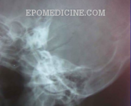
Mastoids XRay is usually ordered by doctors if you have these indications Fever, irritability, and lethargy Swelling of the ear lobeThe classic radiographic assessment of mastoid air cell system size is the Runström II view, but the Law lateral view is the commonly used clinical view in the United States Isolated temporal bone specimens are most accurately positioned using a modified Law Lateral view (with the film perpendicular to the central Xray beam)The Xray mastoid is done to know mastoid pneumatisation and the level of sinus and dural plates Xray mastoids were obtained by Law's view bilaterally and high resolution computed tomography of the temporal bone was obtained with 1mm cuts in axial and coronal planes
A simplified method of producing the axial view of Mayer in chronic mastoiditis and attic cholesteatomaMastoid XRay Diagnostic MRI Mastoid XRay Price Range W Sam Houtson Pkwy South, #150 Houston, TX Work here?Nasal Bone – apl (Water's View And Soft Tissue Lat) Chest Xray (Ap, Lat Below 5 Yrs Old) Nasal Bone Soft Tissue Lat;
Mastoid Abscess Inflamed, tender, and fluctuant right postauricularmastoid swelling Thick abscess drained upon skin incision The abscess cavity was then irrigated with saline and diluted povidine Temporary drain was inserted while continuing antibiotic therapy until significant improvement observed Laws view mastoids positioning" Keyword Found Websites Keywordsuggesttoolcom DA 28 PA 39 MOZ Rank 69 Schuller's view is a lateral radiographic view of skull principally used for viewing mastoid cellsThe central beam of Xrays passes from one side of the head and is at angle of 25° caudad to radiographic plateMastoid Xray (Laws & Mayers) Chest Bucky (Ap) Mastoiditis;
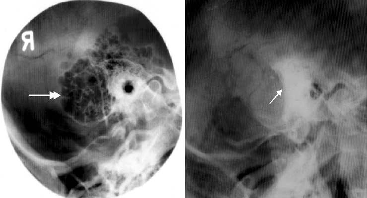


On Radiology Benefits Of Schuller View On Showing Mastoid Bone


Jaypeedigital Ebook Reader
Abstract DURING the past 17 years, I have treated 41 cases of mastoiditis with Xrays Of these, 16 acute cases were seen in children Of the adults, 15 were acute, seven subacute, and three chronic The chief complaints in all were pain, tenderness to pressure over the mastoid Lateral, oblique, anteroposterior, and semiaxial views and modifications of these views were produced by angulation of the xray beam or the patient's head The lateral mastoid view ( Fig 381 ) is the only projection still used in some imaging centers, largely to confirm a diagnosis of acute mastoiditis or substantiate previous mastoid diseaseThis is a panoramic Xray scanning of the mandible and maxilla It is an excellent imaging technique in investigating dental or alveolus related lesions such as odontogenic sinusitis, root canal diseases, ameloblastoma and etc Another example of orthopantomogram Orthopantomogram with braces inplace Further reading Wikipedia


2


Combined Radiographic And Anthropological Approaches To Victim Identification Of Partially Decomposed Or Skeletal Remains Sciencedirect
View this xray 1 Name the view 2 Write down the differential diagnosis Xray both mastoids Laws view (lateral oblique) Differential diagnosis 1 Large antral cell This is usually bilateral 2 Cholesteatomatous cavity Radiologically this cavity will be surrounded by a rim of sclerosis 3 Operated cavity Pt will give h/o mastoidVersion 269 XR Mastoid bilateral Law and Mayer and Stenver and TowneActive FullySpecified Name Component Views Law Mayer Stenver Towne Property Find Time Pt System Head>Mastoidbilateral Scale Doc Method XR Additional Names Short Name XR MastoidBl LawMayerStenverTowne Associated Observations This panel contains the recommended A plain Xray of mastoid/Law's view was done to assess the position of dural and sinus plates Routine haematological and biochemistry investigations were done to assess fitness for anaesthesia After the examination and appropriate investigations informed consent was taken for participation in the trial Those consenting to participate in


2
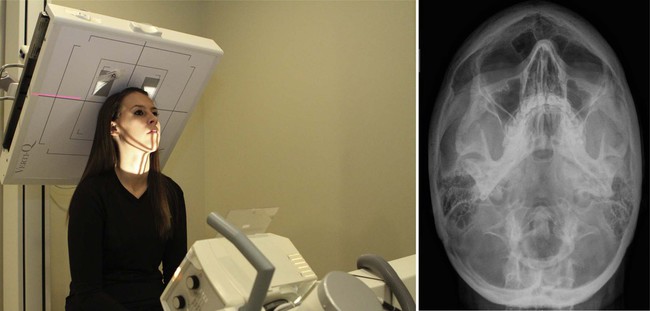


Radiographic Positioning Radiology Key
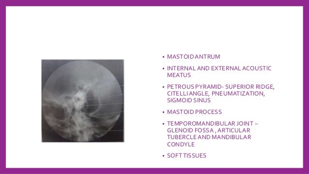
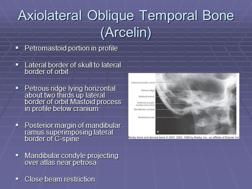
XRay imaging for MASTOID Performed on a Digital XRay Please note that these scans involve XRay radiation, and are not to be performed during pregnancy Test Type Radiology Preparation No Special Preparation Required Reporting Within 24 Hours* Test Price Please choose Location and other options on this page to view final cost in Delhi NCR • mastoid antrum • mastoid air cells • superimposed internal and external acoustic meatuses • mandibular condyle • mastoid process • softtissues 73 petromastoid portion stenvers view • axiolateral oblique posterior profile • prone position, or seatedClinical Discussion Ear Free download as Powerpoint Presentation (ppt / pptx), PDF File (pdf), Text File (txt) or view presentation slides online clinical ear discussion for otologist and audiologist


Xrays In Ent Dr Sujan Chhetri Ms Ent


X Ray Mastoid Lateral Oblique Law S View Left Side Shows Sclerosis Download Scientific Diagram
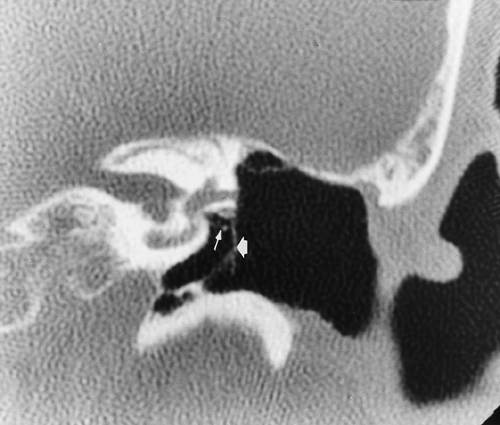
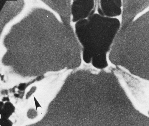
Xray mastoids were obtained by Law's view bilaterally and high resolution computed tomography of the temporal bone was obtained with 1mm cuts in axial and coronal planes Purpose of the study to compare regarding the pneumatisation in chronic suppurative otitis media with xray both mastoids and HRCT temporal boneChest Xray (Each Oblique View) Orbit X We have been studying how to make xray examination of the temporal bone, middle ear, and mastoid process as simple and informative as possible What is required of us by the otologic surgeon is a demonstration of the middle ear and ossicles, the epitympanic space, bony bridge, aditus, and the mastoid antrum


2


2
Procedure for XRay Mastoids (Obl View) Test The process for the Xray oblique view only requires the participation of your upper body The doctor will inject a dye into the area below the infected ear This will help in distinguishing the tissues and nerves clearlyClaim your profile No Reviews Request a Free Quote Diagnostic MRI is a Diagnostic Testing Facility in Houston, TX This medical facility offers procedures at prices which are above average for the market #mastoids#xrayclasseshello!!!!what's up guys, kaise ho dosto?


Jaypeedigital Ebook Reader


60 Radiographs Labeling Ideas Radiography Radiology Technologist Radiology
We also tried to determine the significance of the hereditary and environmental theories of mastoid pneumatization This prospective study consists of 100 patients with unilateral middle ear pathologies over a period of 24 months Bilateral xrays mastoids (law's view) were taken for all the patients The area was measured by using planimetryInfant skull xray lateral view this is an xray image of the skull of an infant taken from a lateral view showing the skull from the side showing 1 frontal bone 2 parietal bones 3 occipital bone 4 lambdoid suture 5 ocular sockets 6 vertex 7 temporal bone 8 mastoid air cells 9 the man Sclerosed mastoid and cavity on xray mastoids and HRCT temporal bone as shown in fig 2 and fig 3 respectively In normal ears, the coincidence of xray and HRCT findings of status of mastoid was 972% except in one case (28%) there was a difference as shown in table 4 In this case it was sclerotic on xray but diploeic on HRCT


Ce4rt X Ray Positioning Of The Mastoid Process For Radiologic Techs


Comparative Medical Radiography Practice And Validation Sciencedirect
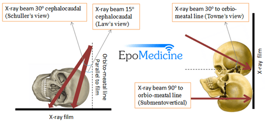
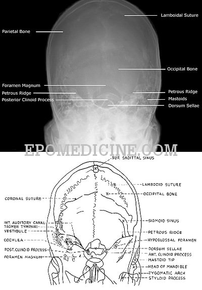
View and Download PowerPoint Presentations on X Ray Mastoid PPT Find PowerPoint Presentations and Slides using the power of XPowerPointcom, find free presentations research about X Ray Mastoid PPTThe xray study of the mastoid region, which was begun in March, 1908, has undergone a slow but gratifying metamorphosisUndertaken with grave doubts as its practical value, it has developed into a method which rivals in its accuracy other recognized methods of physical examinationCentral Ray The horizontal central ray is centered in the midline of the occiput so that the emergent ray exits the patient in the midline at the level of the anterior nasal spine at the upper border of the maxilla Various views for mastoid • LAW's view lateral Oblique view


Digital X Ray Of Mastoid Region Law S Lateral Oblique View Showing Download Scientific Diagram


2
Usually these projection taken in open and closed mouth positions Modified Law method is an xray special projection to best demonstrate the abnormal relationship of temporomandibular fossa or TMJ, which also known as rang of motion between condyles and TM fossa Commonly this projectjion is taken in open and closed mouth positionMastoids XRay may be performed to assess damage to the ear as well as the source of pain and discomfort to the area There are multiple XRay views when checking your mastoids Who should get this test?Before thinsection highresolution CT, many Xray views and modifications were used Today, only few views are used STENVERS VIEW – oblique projection (angled 45° forward) to provide unobstructed view of petrous bone, bony labyrinth, internal auditory canal SCHÜLLER VIEW – along ear canal – demonstrates mastoid air cells
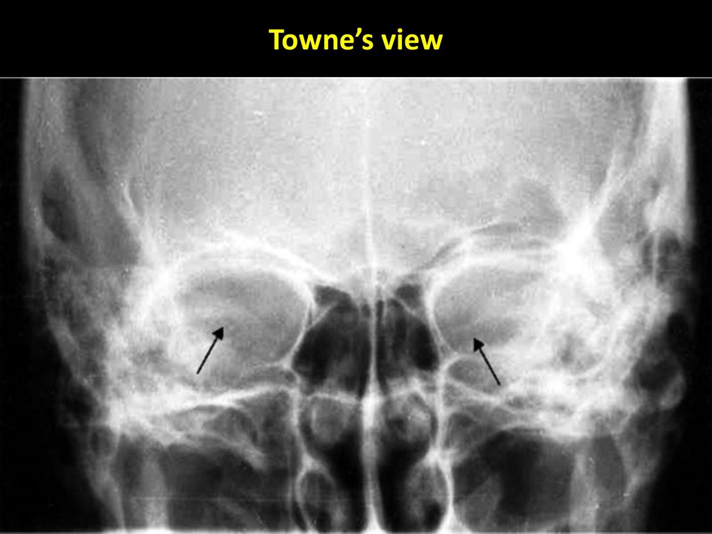


Dr Sujan Chhetri Ms Ent Ppt Video Online Download


Mastoids Lat Obl View Anatomy And Physiology Part 23 Youtube
How to position for mastoid xray Download Here Free HealthCareMagic App to Ask a Doctor All the information, content and live chat provided on the site is intended to be for informational purposes only, and not a substitute for professional or medical adviceInfant skull xray lateral view this is an xray image of the skull of an infant taken from a lateral view showing the skull from the side showing 1 frontal bone 2 parietal bones 3 occipital bone 4 lambdoid suture 5 ocular sockets 6 vertex 7 temporal bone 8 mastoid air cells 9 the man The size of the mastoids was measured by using a graph paper, on which the Xray film of the mastoid taken in the lateral oblique view (Law's view) was superimposed Patients with CSOM (TTD) with less than 3 months of dry ear and small size mastoids on Xray were subjected to cortical mastoidectomy and type I tympanoplasty;


How To Do Mastoids In X Ray Table Youtube


Spring Final Review 15 Flashcards Quizlet
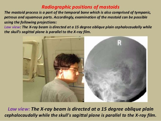
Xray analysis of mastoiditis Radiography of the temporal bone in Cullera is the most appropriate method of roentgenologic examination of its mastoid part Xray detection of bone destructive changes in the initial phase of mastoiditis requires high technical quality radiographs To simplify complex equipment of pictures of the temporal boneDosto aj main app logo ko sikhayunga ki mastoids ka xray app log table pe kaise karenge positi Accordingly, examination of the mastoid can be possible using the following projections Law view The Xray beam is directed at a 15 degree oblique plain cephalocaudally while the skull's sagittal plane is parallel to the Xray film Law view The Xray beam is directed at a 15 degree oblique plain cephalocaudally while the skull's sagittal


Mastoids Radiographic Anatomy Medical Radiography Radiology Imaging Radiology Student
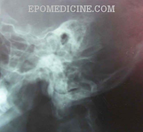


X Ray Of Mastoids Epomedicine
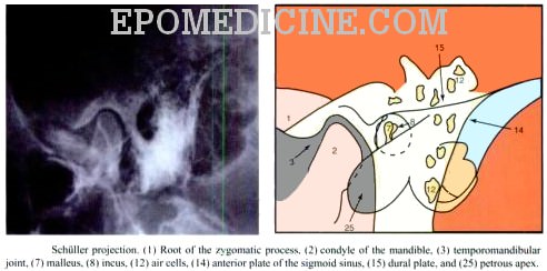
Schüller's view (Runstrom) is a lateral view of the mastoid obtained with the sagittal plane of the skull parallel to the film and with a 30° cephalocaudal angulation of the xray beam These 30° in Schüller's view displaces the arcuate eminence of the petrous bone downward and shows the antrum and the upper part of the atticSchuller's view is a lateral radiographic view of skull principally used for viewing mastoid cells The central beam of Xrays passes from one side of the head and is at angle of 25° caudad to radiographic plate This angulation prevents overlap of images of two mastoid bones Radiograph for each mastoid is taken separatelyThe size of the mastoids was measured by using a graph paper, on which the Xray film of the mastoid taken in the lateral oblique view (Law 's view) was superimposed Patients with CSOM (TTD) with less than 3 months of dry ear and small size mastoids on Xray were subjected to cortical mastoidectomy and type I tympanoplasty;


Jaypeedigital Ebook Reader


Mastoid Series Normal Radiology Case Radiopaedia Org
Mastoid process is the conical prominence that extends from the temporal bone, just behind where the ear is locatedThe neck muscles are attached to the mastoid process It is filled with cavitiesAxiolateral ObliqueModified Law Method CENTRAL RAY CR directed to a midpoint of the grid at an angle of 15 degrees caudad to exit the downside mastoid tip approximately 1 inch posterior to the EAM The CR enters approximately 2 inches posterior to, and 2 inches superior to the uppermost EAMThe modified Stenvers view is an oblique radiographic projection used to demonstrate the petrous temporal bone, IAM and bony labyrinthIt is performed as a posteroanterior (PA) projection to minimize radiation to the orbits This view has succeeded the Stenvers view, which includes more of the mastoid air cells


1


2


Spring Final Review 15 Flashcards Quizlet


Mastoid Series Normal Radiology Case Radiopaedia Org


X Ray Mastoid Lateral Oblique Law S View Left Side Shows Sclerosis Download Scientific Diagram


2


Digital X Ray Of Mastoid Region Law S Lateral Oblique View Showing Download Scientific Diagram


2


Mastoids Lat Obl View Anatomy And Physiology Part 23 Youtube


2
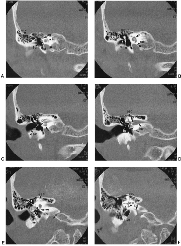


The Temporal Bone Radiology Key
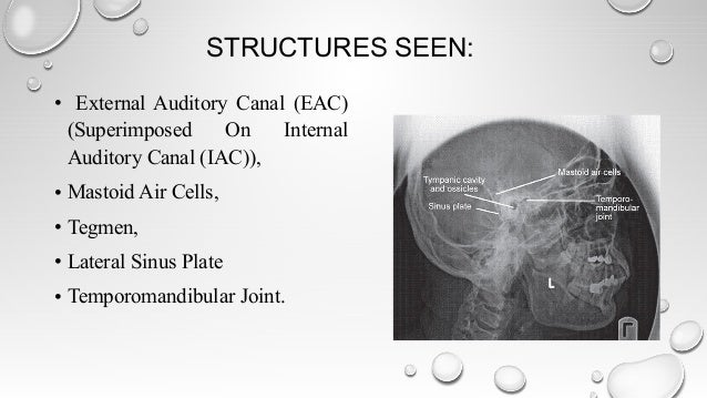


Radiological Imaging In Head And Neck And Relevant Anatomy


Stenvers View Radiology Reference Article Radiopaedia Org
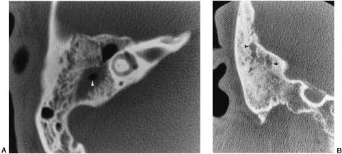


The Temporal Bone Radiology Key


Q Tbn And9gctj3zr Hrj2dnckcxuveb Enkdddksnlfto1uq1smrpcw7sbym2 Usqp Cau


2


Osce Notes In Otoradiology By Drtbalu Osce Notes In Otolaryngology


X Ray Of Mastoids Epomedicine


Mastoid Stenvers View Youtube


Jaypeedigital Ebook Reader


X Ray Of Mastoids Epomedicine


2


Pps Radiology


2


Ce4rt X Ray Positioning Of The Mastoid Process For Radiologic Techs


Jaypeedigital Ebook Reader


X Ray Mastoid Lateral Oblique Law S View Left Side Shows Sclerosis Download Scientific Diagram


2


X Rays In Ent
:background_color(FFFFFF):format(jpeg)/images/library/12296/chest_PA.jpg)


Radiological Anatomy X Ray Ct Mri Kenhub


X Ray Of Mastoids Epomedicine


Radiographic Positions Of Mastoids Human Head And Neck Human Anatomy


Mastoid Stenvers View Youtube


X Ray Of Mastoids Epomedicine


Mastoid Stenvers View Youtube
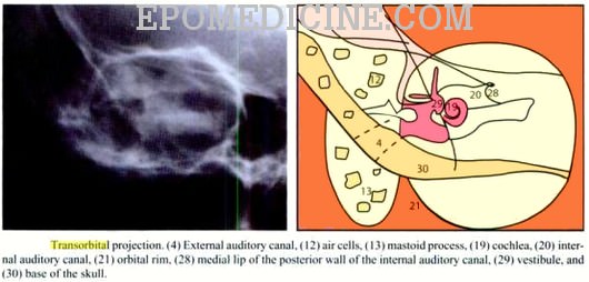


X Ray Of Mastoids Epomedicine


2


Schuller S View Wikipedia
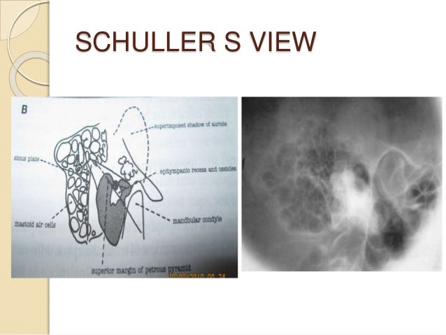


Radiology In Head And Neck By Kanato T Assumi


2


2


Right Well Developed Aerated Mastoid Lateral X Ray Download Scientific Diagram
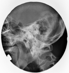


스크랩 Mastoid Law View
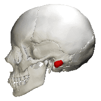


Ce4rt X Ray Positioning Of The Mastoid Process For Radiologic Techs


Q Tbn And9gcsbrmx1mjyuc2bv61h6flzmqtzn H4el8pg 0fr0kg Usqp Cau


X Ray Of Mastoids Epomedicine


The Temporal Bone Radiology Key


Presentation1 Pptx Radiological Anatomy Of The Petrous Bone


View Image


The Temporal Bone Radiology Key


2
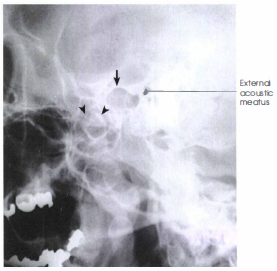


Modified Law Method Tmj Radtechonduty
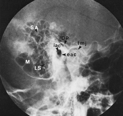


The Temporal Bone Radiology Key


2
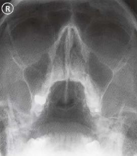


Ce4rt Radiographic Positioning Face And Mandible For X Ray Techs
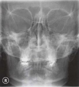


Ce4rt Radiographic Positioning Face And Mandible For X Ray Techs


Q Tbn And9gcruyyv Hw1o R9t1wsych6rjpkvu4n2dz8ln9gndramxi4rvlk5 Usqp Cau


2


Jaypeedigital Ebook Reader


Pns Pns Water View Positioning Anatomy And Physiology Part 21 Youtube


X Ray Of Mastoids Epomedicine


Skull Radiography Techniques And Reporting


Radiology In The Study Of Bone Physiology Academic Radiology


A And B X Rays Both Mastoids Law S View Showing Radio Opaque Foreign Download Scientific Diagram


Mastoids And Organs Of Hearing Ppt Video Online Download


60 Radiographs Labeling Ideas Radiography Radiology Technologist Radiology


2


Cochlear Implant Radiology Reference Article Radiopaedia Org


X Ray Of Mastoids Epomedicine
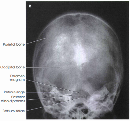


Skull Towne Method Ap Axial Projection Radtechonduty


2


Comparative Medical Radiography Practice And Validation Sciencedirect


Skull Towne View Radiology Reference Article Radiopaedia Org


Mastoids Radiographic Anatomy Wikiradiography


Top Photos In Infant Skull X Ray Lateral View


Role Of X Rays In Otolaryngolgoy Esophagus Medical Imaging
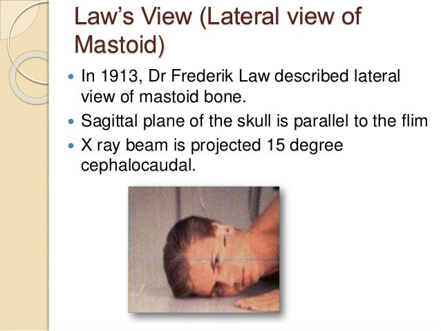


Radiology In Head And Neck By Kanato T Assumi
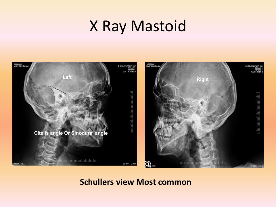


Laws View X Ray 鬼画像無料


Digital X Ray Of Mastoid Region Law S Lateral Oblique View Showing Download Scientific Diagram


2


